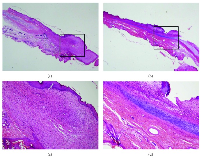
Figure 4.
Histology of auricular cartilage defect. (a) Normal cartilaginous tissue was not visible in black square box in the control group. (b) New cartilage tissue was shown in black square box in the experimental group. (c) Fibrous tissue formation was observed instead of chondrocyte and extracellular matrix (ECM). (d) Typical cartilaginous features with chondrocytes, chondroblasts, and cartilage-specific ECM deposition were shown. Sections were stained with hematoxylin and eosin (magnification, a, b: 10x, c, d: 200x).

