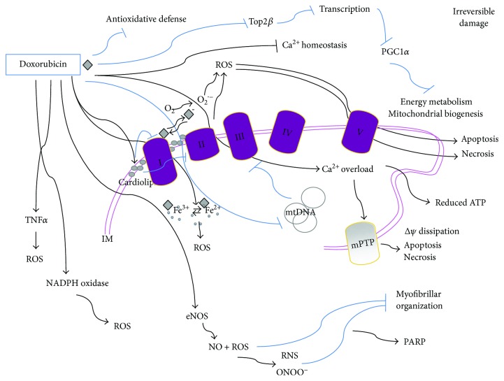
Figure 1.
Doxorubicin-mediated cytotoxicity is mostly irreversible. Mitochondrial doxorubicin accumulation is due to its specific binding to the phospholipid cardiolipin; this membrane perturbation inhibits complex I and complex II disrupting the electron transport chain and inducing ROS production. ROS might also be produced by other doxorubicin-mediated mechanisms: a quinone moiety in the chemical structure of doxorubicin is reduced by complex I into a reactive semiquinone free radical which transfers an electron to O2 and generates the superoxide anion O2•−. In turn, the semiquinone free radical is oxidized and returns to the quinone form in a sequence of reactions known as the “redox cycling” of doxorubicin. Moreover, doxorubicin can directly interact with iron to form reactive anthracycline-iron complexes resulting in an iron cycling between Fe3+ and Fe2+ associated with ROS production and altering iron homeostasis. Doxorubicin also induces mtDNA damage and binds to eNOS enhancing its activity thus leading to NO production and contributing to peroxynitrite (ONOO−) formation. It also disrupts Ca2+ homeostasis which triggers mPTP and dissipates the transmembrane potential (ΔΨ) along with increasing mitochondrial permeability to apoptotic factors such as cytochrome c and leading to apoptosis or necrosis. The excessive oxidative stress produced by doxorubicin can also be mediated by increasing levels of TNFα and by NADPH oxidase and leads to redox modifications of macromolecules such as myofibrillar proteins. Doxorubicin also reduces the antioxidative defense of cells, and by preventing Top2β activity, it alters the transcriptome, for example, downregulating PGC-1α, which negatively impacts on both oxidative phosphorylation and mitochondrial biogenesis. IM: inner membrane space.

