
Figure 2.
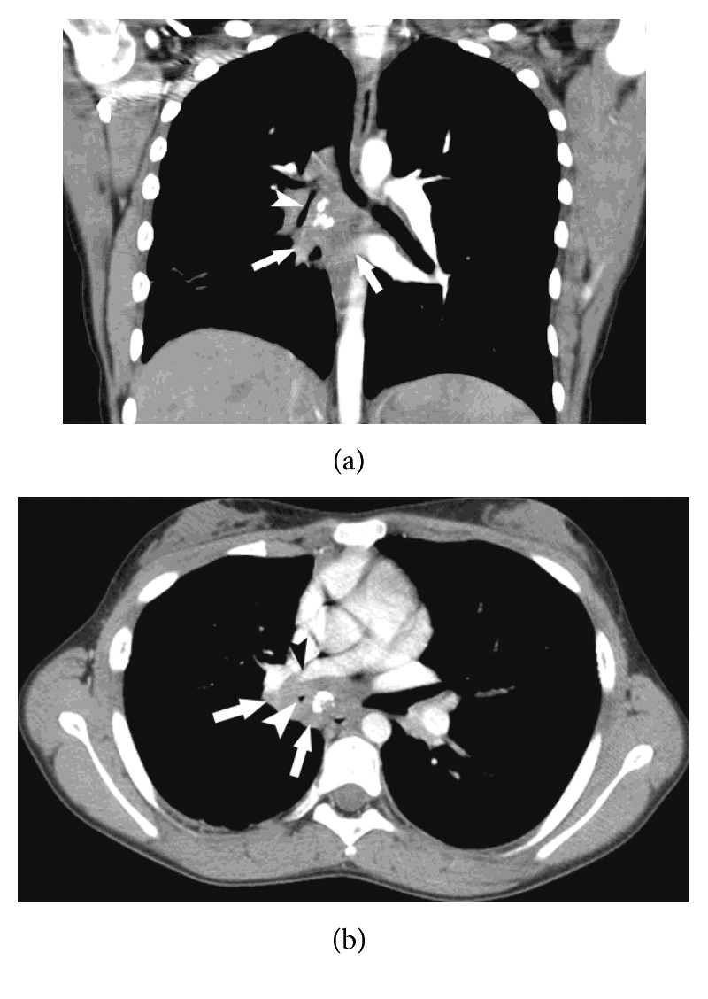
Axial (a) and coronal (b) reformats of the chest CT following a contrast injection, showing a subcarinal and right hilar ill-defined soft tissue mass (white arrows) with prominent interior calcifications causing marked compression on the bronchus intermedius, right middle lobe bronchus (white arrowhead), and right inferior pulmonary vein (black arrowhead).