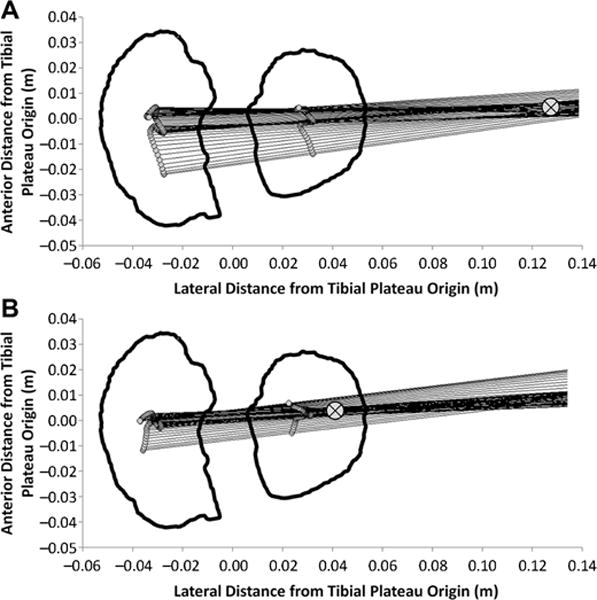
Figure 1.

Medial-lateral axis of the femur projected onto the transverse plane of the tibial plateau is shown at each instant during the stance phase of walking for a characteristic participant with a highly lateral KCOR at (A) 2 years after ACLR and (B) the same participant at 4 years after ACLR with a more medial KCOR. ⊗ indicates the location of KCOR, which was calculated as the least squares intersection of the projected lines. Video versions of Figure 1A and 1B showing the time history throughout the stance phase of the medial-lateral axis of the femur can be found in the Video Supplement. ACLR, anterior cruciate ligament reconstruction; KCOR, knee center of rotation.
