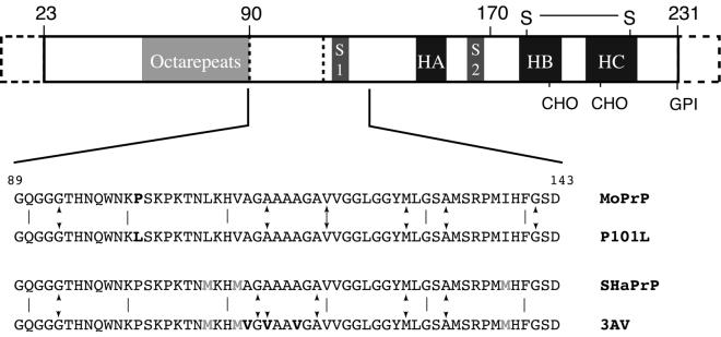
Figure 1.
A schematic diagram of the PrP showing the sequence motifs and secondary structure of PrPC. The segment marked octarepeats contains six copies of an eight-residue repeat sequence, S1 and S2 are the two small β-strands, and HA, HB, and HC designate the three helices. Residues 1–23 are processed off during transport. The points marked CHO indicate the sites at which oligosaccharides are attached in vivo, and GPI indicates the attachment point of the membrane anchor. The region 23–124 is unstructured in PrPC in solution. Below the sequences of peptides studied are given, and the sites of sequences variations are highlighted. Sites of incorporation of isotope labels are indicated by arrows.

