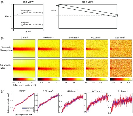
Fig. 6.
Multispatial frequency reflectance results obtained on a phantom containing a slanted absorbing tube ranging in depth from 0 to 5 mm, containing an absorbing dye with a scattering background. (a) Schematic of phantom geometry. (b) Reflectance maps calibrated to a homogenous tissue-simulating phantom are derived for DC (), 0.06, 0.09, 0.12, and using the fundamental and second harmonic components from two square wave patterns. (c) Cross-sections of calibrated reflectance taken from horizontal line in center of image where tube is located. Mean reflectance values along the line agree to within 0.04%, 0.1%, 0.4%, 0.1%, and 0.12% for 0, 0.06, 0.09, 0.12, and respectively.

