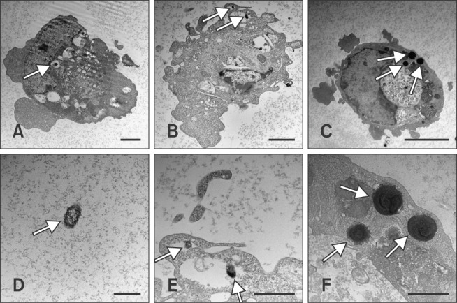
Fig. 3. Micrographs of Pasteurella multocida B:2 infected dexamethasone-, lipopolysaccharide-treated, and untreated bovine aortic endothelial cells (BAECs). The cells were treated with 10 µM dexamethasone (B) or 100 ng/mL lipopolysaccharide (C) for 24 h, and the treated cells were incubated with P. multocida B:2 for 2 h. The untreated cells (A) were used as the control and (D) showed the P. multocida B:2. Micrographs (E) and (F) are enlargements of micrographs (B) and (C), respectively. The white arrows indicate a bacterium. The bacteria resided in vacuolar compartment of dexamethasone-treated and untreated cells, whereas the bacteria resided within the plasma membrane of lipopolysaccharide-treated cells. The samples were observed under transmission electron microscope (H-7100; Hitachi) operating at 100 kV. Scale bars = 1 µm (D-F); 2 µm (A and B); 5 µm (C).

