Figure 2.
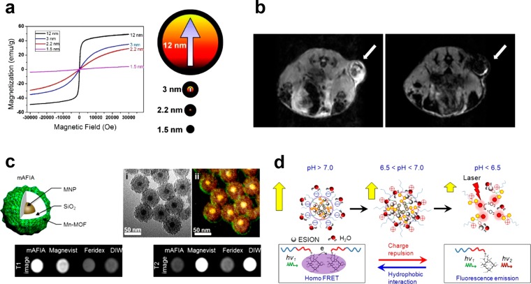
(a) Field-dependent magnetization (M–H) curves at 300 K for iron oxide nanoparticles of various sizes (left). Description of the spin canting effect in the iron oxide nanoparticles of various sizes (right). (b) In vivo MR images of the xenografted tumor before (left) and after (right) intravenous administration of FIONs. (c) Artifact filtering imaging agent (mAFIA) that comprises a combination of paramagnetic Gd-MOF and superparamagnetic nanoparticles for T1–T2 dual-mode MRI. (d) Tumor pH-responsive magnetic nanogrenades composed of self-assembled extremely small-sized iron oxide nanoparticles and pH-sensitive ligands. Reprinted with permission from refs (21, 38, 67), and (74). Copyright 2011, 2012, and 2014 American Chemical Society.

