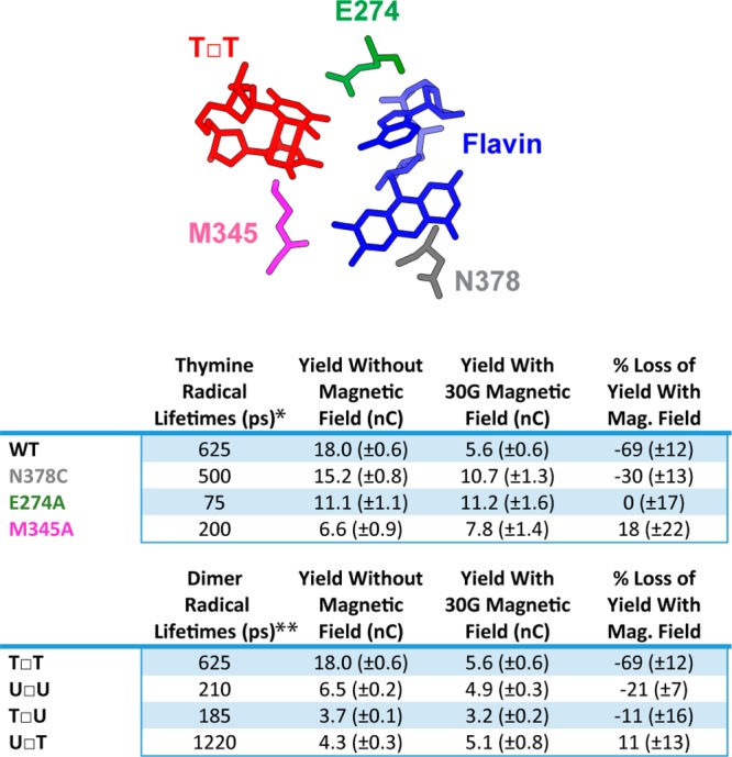
Figure 6.

Effect of structural perturbations on magnetosensitivity. (Top) Cartoon showing placement of thymine dimer relative to flavin cofactor and three active site residues in photolyase. Positions of residues adapted from DNA-bound photolyase crystal structure.15 (Middle) Comparison of the yield of charge transferred to different active site mutants with and without 30 G magnetic field perpendicularly intersecting the plane of the electrode surface after 60 min of irradiation with blue light anaerobically in Tris buffer (50 mM Tris-HCl, 50 mM KCl, 1 mM EDTA, 10% glycerol, pH 7.5). (Bottom) Comparison of the yield of charge transferred to different cyclobutane pyrimidine dimers with and without 30 G magnetic field perpendicularly intersecting the plane of the electrode surface after 60 min of irradiation with blue light. For these dimers U = uracil, T = thymine, and the 5′ position is listed first with the 3′ position second. Standard error was included with n ≥ 6. *Lifetimes of thymine radicals with mutant photolyase were obtained from C. Tan et al.13 **Lifetimes of different dimer radicals were obtained from Z. Liu et al.14
