
Fig. 2.
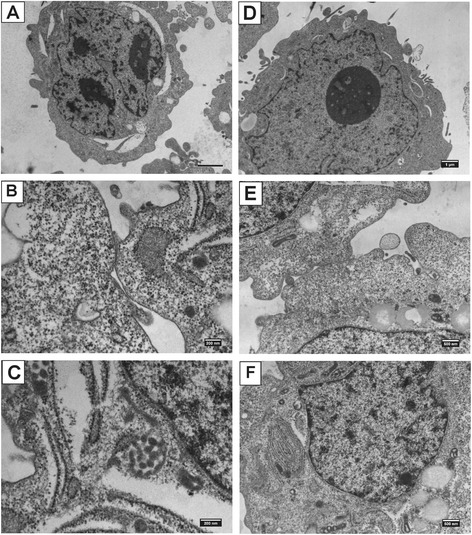
Representative images of electron microscopy of RSBS-14 (a-c) and RSBS-43 (d-f) derived from squamous cell carcinoma and adenocarcinoma respectively. RSBS-14- a High nucleus to cytoplasmic ratio, b Hemidesmosomes, c Keratin filaments. RSBS-43- d Prominent nucleolus, e Cell junctions f Prominent rough endoplasmic reticulum