Fig. 4.

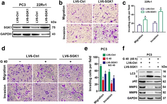
SGK1 overexpression attenuates autophagy and promotes cell migration and invasion. a-c PC3 and 22Rv1cells were infected with lentivirus containing an SGK1 (LV6-SGK1) expression plasmid or an empty vector (LV6-Ctrl) that confers resistance to puromycin. PC3 and 22Rv1cells cells stably overexpressing SGK1 or vector alone were assessed using a western blot against SGK1 (a), 6 × 10⁴ 22Rv1 for migration, 3 × 10⁵ 22Rv1 for invasion (b). Representative histograph of invaded tumor cells is displayed and number of invaded tumor cells quantified (c). d-f PC3 cells stably overexpressing SGK1 or vector alone were treated for 24 h with 40 μM GSK650394. Cell migration assays and matrigel cell invasion assays were performed. Representative fields of migration and invasion cells on the membrane are shown (magnifications, × 200) (d). And number of invaded tumor cells per field is quantified (e). Total protein lysates were collected after 48 h treatment with 40 μM GSK650394 for western blot against LC3, p62, MMP3 and MMP9, and GAPDH was used as a loading control (f). All results are representative of three experiments and are expressed as the mean ± S.D.; *P < 0.05
