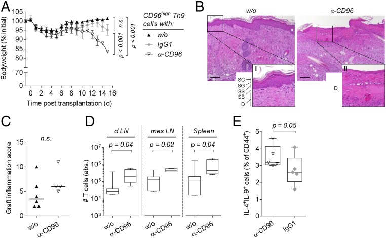
Fig. 6.
Blockade of CD96 in CD96high Th9 cells restores their inflammatory capacity. (A–D) C57BL/6 Rag1−/− mice were reconstituted with 1 × 10⁵ CD96high cells sorted from alloreactive C57BL/6 Th9 cultures and received i.p. injection of 250 µg of anti-CD96 (α-CD96, n = 4) or isotype control (IgG1, n = 3) antibody or were left untreated (w/o, n = 6–7). All animals received a BALB/c skin graft on the next day. (A) Body weight of mice shown as mean ± SEM. P value of the interaction term (group with time) between two groups was calculated using an ANOVA type III test after fitting a linear mixed-effect model to the data. (B) Representative hematoxylin and eosin stained sections of skin grafts harvested 10 d post skin transplantation. Higher magnification of w/o photograph (I) displays the epidermis composed of the stratum corneum (SC), stratum granulosum (SG), stratum spinosum (SS), stratum basale (SB), and part of the dermis (D). Magnification of α-CD96 photograph (II) shows the altered dermis without epidermis. (C) Inflammation score of skin grafts harvested between day 9 and 15 posttransplantation. Median is shown, and a one-tailed Mann–Whitney test was applied. (D) Absolute number of T cells isolated from graft draining lymph nodes (d LN), mesenteric lymph nodes (mes LN), and spleen harvested 9 to 15 d posttransplantation. P values were calculated with a two-tailed Mann–Whitney test. (E) Frequency of IL-4+IL-9+ double-producing cells in alloreactive C57BL/6 Th9 cultures following treatment with 5 µg/mL anti-CD96 (α-CD96, n = 5) or isotype control antibody (IgG1, n = 5). Antibodies were added after 24 h of cocultivation, and intracellular cytokine expression was analyzed following an additional 48 h of culture. A one-tailed Mann–Whitney test was applied. n.s., not significant (P > 0.05).

