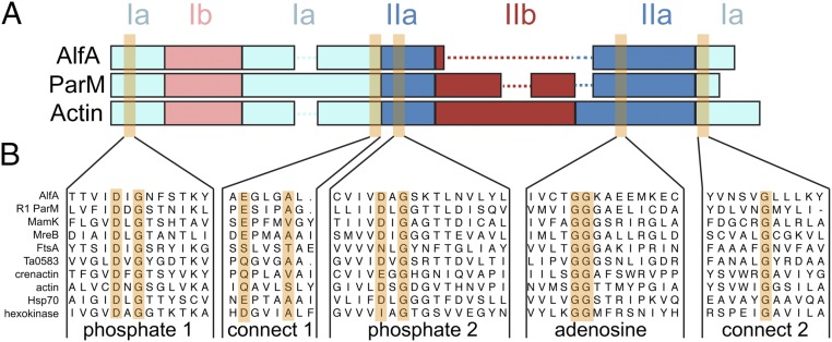
Fig. 1.
AlfA sequence conservation. (A) Diagram of domain arrangements for AlfA, the bacterial actin ParM from the E. coli R1 plasmid, and vertebrate actin. The five conserved actin sequence motifs surrounding the actin binding cleft are highlighted in orange. (B) Sequence alignments of AlfA with other actins, Hsp70, and hexokinase in the regions surrounding the five conserved motifs.

