Fig. 5.
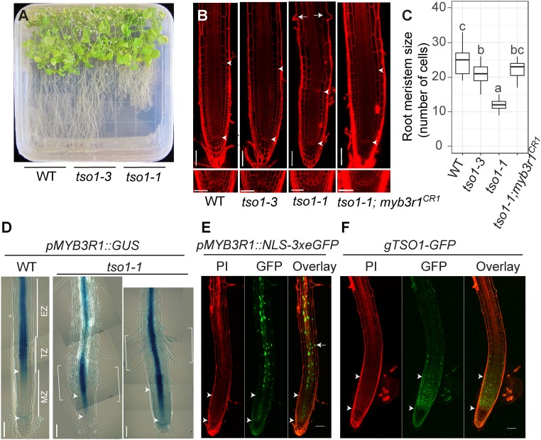
The tso1-1 short root phenotype is suppressed by myb3r1 mutations. (A) WT (Ler), tso1-3, and tso1-1 seedlings on a half strength MS media plate at 42 d postgermination (dpg). (B) Confocal images of root apical meristem of WT (Ler), tso1-3, tso1-1, and tso1-1; myb3r1CR1 at 7 dpg. The Upper arrowhead marks the upper boundary of the MZ as it marks the first cortex cell that is twice the size of the previous cell (cortex cell layer is the second from the epidermis). The Lower arrowhead marks the lower boundary of the MZ as well as the position of quiescent center (QC). Early differentiation of tso1-1 root is indicated by precocious formation of root hairs (arrows). Lower shows the stem cell niche in the root of respective genotype. [Scale bars, 50 μm (Upper) and 25 μm (Lower).] (C) Quantification of root MZ size by the cell number in cortex file in MZ (n = 10–15 roots). Different letters indicate statistically significant difference (P < 0.01, one-way ANOVA and Tukey test). (D) pMYB3R1::GUS expression in wild-type and tso1-1 root at 7 dpg. The MZ near the root tip is followed by the TZ, and then the EZ, which is shown by several images joined together with each image capturing a segment of the root. GUS signal, which reflects the promoter activity of MYB3R1, is restricted to the stele in the TZ and EZ. In tso1-1 mutant, the bracketed segment showing strong GUS signal is shifted downward. The region of ectopic MYB3R1 expression correlated with dense root hair formation. (E) In pMYB3R1::NLS-3xeGFP WT plant, nuclear GFP signals are found in the stele of the root in the EZ and the TZ, consistent with the GUS signal in D. (F) In WT plants containing the gTSO1-GFP construct, nuclear TSO1-GFP is largely restricted in the MZ and appears complementary to the MYB3R1 expression zone. Propidium iodide (PI) staining gives the red outline of the root cells. [Scale bars, 50 μm (B, E, and F) and 100 μm (D).]

