Fig. 3.
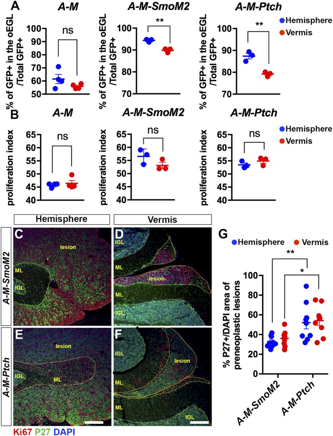
GCPs located in the hemispheres are more sensitive to elevated HH signaling, which maintains them in an undifferentiated state. (A and B) Graphs of the proportions of undifferentiated GCPs (GFP+ in the proliferating outer EGL/total GFP+ cells) (A) and the proliferation index (percent GFP+ EdU+ cells/GFP+ in outer EGL) (B) in H and V of P8 A-M (n = 4), A-M-SmoM2 (n = 3), and A-M-Ptch (n = 3) mice. Significances determined using paired Student’s t test to compare H and V within an animal of each genotype. (C–F) Flourescent immunohistochemical (FIHC) detection of Ki67, P27, and DAPI on sagittal sections of P21 A-M-SmoM2 and A-M-Ptch mice. Internal granule cell layer (IGL), molecular layer (ML), and lesions are indicated with yellow dotted lines. (Scale bars, 200 μm.) (G) Quantification of cell differentiation (P27+ over DAPI+ area) in preneoplastic lesions located in H and V of P21 A-M-SmoM2 (n = 3) and A-M-Ptch (n = 3) mice. Significances determined using two-way ANOVA (overall P = 0.0001) followed by a Sidak post hoc test. All data are expressed as mean ± SEM. **P < 0.01, *P < 0.05, ns, nonsignificant. Statistics are provided in Table S2.

