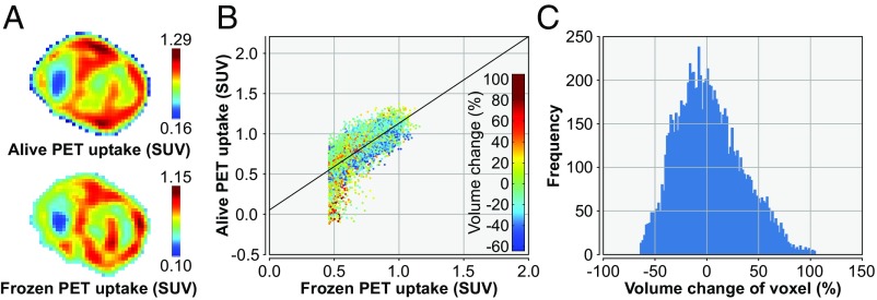
Fig. 4.
An example of the alignment of the PET scans acquired pre- and postfreezing. (A) Frozen PET (Lower) and aligned prefreezing PET (Upper). (B) Scatter plot of the voxel values from the frozen PET (horizontal axis) and the aligned prefreezing PET (vertical axis), with color indicating the amount of deformation required. (C) Histogram of the required change in voxel size for proper alignment.

