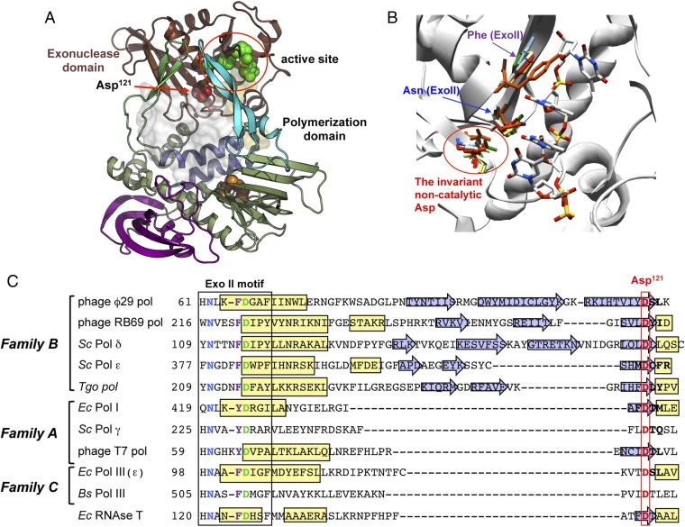
Fig. 1.
(A) Structural view of ϕ29 DNAP and Asp121 location. Cartoon representation of ϕ29 DNAP [Protein Data Bank (PDB) code 2PYJ]. The color code is brown and green for the exonuclease and polymerase domains, respectively. Subdomains in polymerase domain are highlighted in different colors (TPR1 and TPR2 regions in purple and lime-green colors, respectively; fingers in blue; and thumb in cyan). The Asp121 loop is highlighted in red, with Asp121 being shown as spheres. Catalytic residues for exonuclease and polymerase activities are shown as green and orange spheres, respectively. DNA locations are shown by transparent surface representation, in light orange for ssDNA (PDB ID code 2PY5) and in white for primer template DNA (PDB ID code 2PYJ). (B) Structural superposition of the exonuclease active site of ϕ29 DNAP (PDB ID code 1XHX), RB69 DNAP (PDB ID code 2P5G), S. cerevisiae Pol δ (PDB ID code code 3IAY) and ε (PDB ID code 4M8O), T. gorgonarius DNAP (PDB ID code 2XHB), E. coli DNAP I (PDB ID code 1DPI), T7 DNAP (PDB ID code 1TK8), and E. coli DNAP III (PDB ID code 2HNH). (C) Multiple sequence alignment of the Exo II motif and the noncatalytic aspartate in family A, B, and C members. DNAP sequence references were compiled by Braithwaite and Ito (34), with the exception of bacteriophage DNAP RB69 (National Center for Biotechnology Information reference sequence YP_009100595.1); S. cerevisiae (Sc) Pol ε (GenBank accession no. AJT09723.1); T. gorgonarius (Tgo) DNAP (NCBI reference sequence WP_088885078.1); Sc Pol γ (GenBank accession no. 854508); and RNase T of E. coli (Ec) (NCBI reference sequence WP_074465944.1), which belongs to the DNAQ-like superfamily. The α-helices are represented in yellow, and the β-sheets are indicated with blue arrows. The conserved aspartate of the Exo II motif and the noncatalytic aspartate are shown in green and red, respectively, and the conserved asparagine and aromatic residue (phenylalanine or tyrosine) from the Exo II motif are colored in blue and purple, respectively. Bs, B. subtilis.

