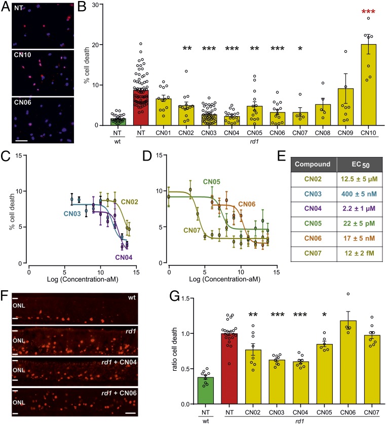
Fig. 2.
Screening of cGMP analogs in vitro. cGMP analogs were tested in retinal neurosphere-derived rod photoreceptor-like cell cultures (A–E) and organotypic retinal explant cultures (F and G). NT, not treated. (A) rd1 mutant photoreceptor-like cell cultures treated with 50 µM of the respective compound until DIV11 (red, ethidium homodimer staining; blue, nuclear counter staining). (B) Percentage of ethidium homodimer-labeled, dying photoreceptor-like cells [green, WT; red, rd1; yellow, different analog treatments; n = 5–62 cell culture samples obtained from two to seven independent preparations; red asterisks indicate toxic effect (i.e., more dying cells compared with NT)]. (C and D) Dose–response curves for CN02–CN07 starting at attomolar concentration. (E) EC50 values for CN02–CN07 calculated from dose–response curves. (F) TUNEL assay (red) on vertical sections from P11 retinal cultures NT or treated with 50 µM of the respective compound. (G) Ratio (treated/NT) of TUNEL-labeled cells (same color code as in B, WT, n = 9 separate retinal explant cultures; rd1: NT, 22; CN02, 8; CN03, 8; CN04, 8; CN05, 6; CN06, 4; CN07, 8). Error bars: SEM. Statistical comparisons: B and G, NT-rd1 vs. treated rd1 using one-way ANOVA with Holm–Šidak’s multiple-comparisons test; ONL, outer nuclear layer containing rod and cone photoreceptor nuclei. (Scale bars: A, 100 µm; F, 50 µm.) *P ≤ 0.05, **P ≤ 0.01, ***P ≤ 0.001.

