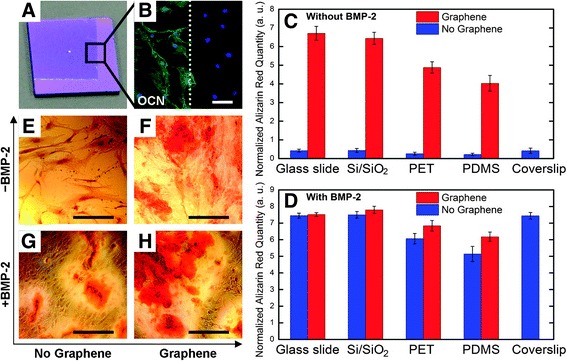
Fig. 2.

Enhancement of osteogenic differentiation on graphene substrates with/without BMP-2. (a) Optical image of graphene-coated Si/SiO2 substrate. The boundary is shown for the graphene-coated part. (b) Osteocalcin (OCN) staining, a marker of osteogenic differentiation. Green = OCN, Blue = DAPI. (c, d) Alizarin Red S (ARS) quantification graphs during 15 days on substrates with/without graphene. (e-h) polyethylene terephthalate (PET) substrate stained with ARS, showing calcium deposits due to osteogenic differentiation. Reprinted with permission from [11]. Copyright (2011) American Chemical Society
