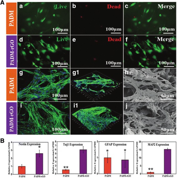
Fig. 7.

The effects of 3D porcine acellular dermal matrix (PADM) and PADM-reduced graphene oxide (PADM-rGO) on the adhesion and neuronal differentiation of human mesenchymal stem cells (hMSCs). (a) The cytocompatibilities of the two different scaffolds. The hMSCs were cultured on the PADM (a, b, c) and PADM–rGO (d, e, f) for 24 h, Live/dead staining was performed. The live cells are stained green, and dead cells are red. CLSM fluorescence morphologies of the actin cytoskeleton of the hMSCs cultured on the PADM (g) and PADM–rGO (i) scaffolds for 3 days. (h – j) SEM images represent the cell attachment of hMSCs after 3 days on the PADM and PADM-rGO. (b) Quantification of qPCR analysis for neural marker genes; Nestin, Tuj1, GFAP, and MAP2, expression of hMSCs. Copyright © 2015, Royal Society of Chemistry
