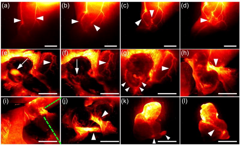
Figure 5. Intra-operative assessment of the vascular supply using NIR-II CQS1000 for precise tumor resection.
(a)–(d). Pre-evaluation of the blood supply of the tumor. White arrowheads indicate the major blood vessels to supply the tumor. (e) and (f) Intra-operative fluorescent images, indicating that the sentinel lymph node(iliac lymph node) and the concomitant artery were easily exposed and identified (white arrows), respectively. White arrowheads indicate the major blood vessels to supply the tumor. (g) and (h). Major blood vessels supporting the tumor were dissected and exposed (white arrowheads). Major blood vessels were further dissected surgically into two small branches (i) and magnified as (j). As the ex vivo image shown in (k), the major blood artery and vein of tumor were successfully ligated (white arrowheads). Scale bar: 4mm.

