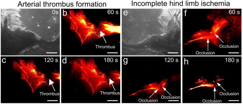
Figure 6. In vivo NIR-II images for arterial thrombus formation and incomplete hind limb ischemia.
NIR-II images of the left hind limb were captured at 0s (a), 60s (b), 120s (c), 180s (d) after CQS1000 injection intravenously into the tail vein of C57BL/6 mice. White arrows indicate the thrombus formation in femoral artery. NIR-II images of the left hind limb were captured at 0s (e), 60s (f), 120s (g), 180s (h) after CQS1000 injection intravenously into the tail vein of C57BL/6 mice. White arrows indicate the incomplete ischemia (occlusion) in femoral artery. Scale bar: 4mm.

