
Fig. 5.
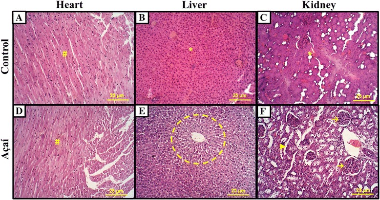
Açaí reduce toxicity effects in breast cancer experimental. Microscopic analysis: (a, d) no morphological difference in heart was observed with presence of cardiac muscle tissue well preserved (#). (b, e) In açaí treatment, we note the presence of centrilobular vein and cords of hepatocyte (circle) showed normal liver tissue. However, we observed higher toxicity effects in control liver with higher fibrosis, atypical cells and hemorrhagic microenvironment (*). (c, f) Similarly, in kidney analysis we also demonstrated increased toxicity in control (*), while in the açaí group, we observed the presence of numerous glomeruli (→) and apparent distal tubules (►)