
Figure 5.
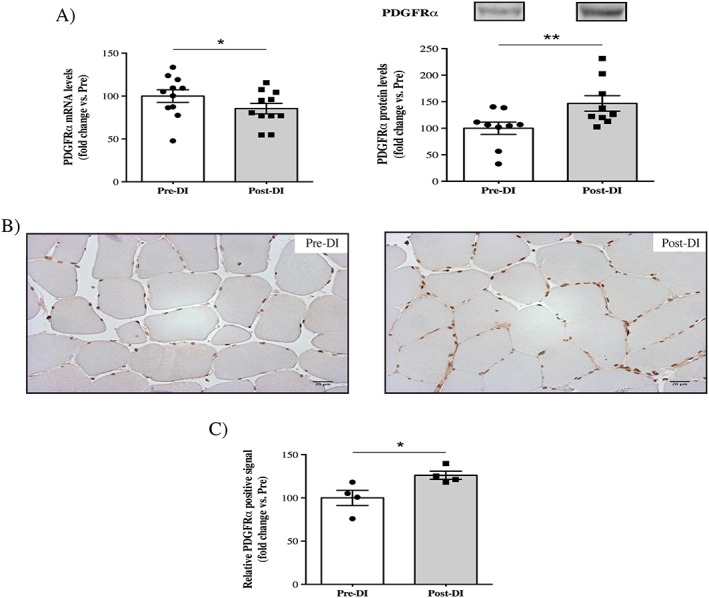
Changes in the fibro‐adipogenic progenitor cell surface marker PDGFRα after 3 days of dry immersion. (A) Changes in PDGFRα mRNA and protein levels in vastus lateralis muscle biopsies taken before (Pre‐DI) and after (Post‐DI) 3 days of dry immersion (DI). (B) Representative histological transversal paraffin‐embedded vastus lateralis muscle sections that were taken from Pre‐DI and Post‐DI muscle biopsies are immunostained with PDGFRα antibody. (C) Quantification of the PDGFRα‐positive signals. * P < 0.05 and ** P < 0.01.