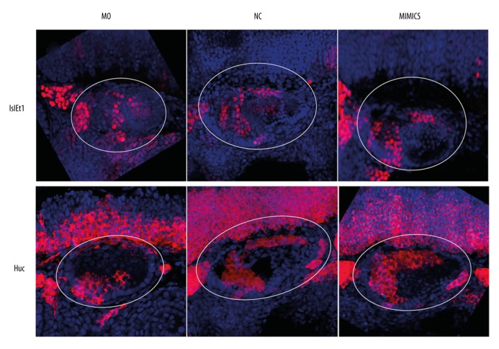
Figure 6.
MiR-194 promoted the development and differentiation and regulation of the normal spatial structure of SAG in zebrafish inner ear. The percentage of Islet1 (upper portion) and HuC (lower portion) were measured by in situ hybridization at 48 hpf. The white circle area indicated zebrafish inner ear. Red staining indicated Islet1 expression (upper portion) or HuC expression (lower portion); blue indicated cell nucleus. MO – microinjection with morpholino oligonucleotide; NC – negative control; Mimics – microinjection with miR-194 mimics; hpf – hour post fertilization; SAG – statoacoustic ganglion.

