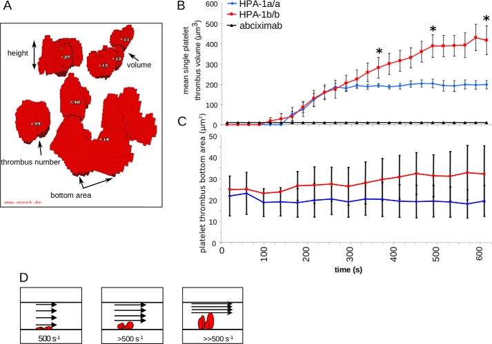
Figure 2.
Dynamics and volumetric analysis of platelet thrombus formation under flow-dynamic conditions. A rectangular flow chamber coated with collagen type I (3 mg/ml) at the lower surface was perfused with mepacrine-labeled citrated whole blood for 10 min at an initial near-wall shear rate of 500 s−1, simulating arterial flow conditions. Fluorescence signals were detected by confocal laser scanning microscopy, and digital imaging was processed as described under “Experimental procedures”. Volumetry of forming platelet thrombi was assessed by real-time 3D visualization. A, a reconstruction of formed platelet thrombi obtained from a stack of 30 images by confocal laser scanning microscopy and subsequent data processing by ECCET. B and C, initial platelet thrombus formation and subsequent thrombus growth were recorded in 25-s intervals for each single thrombus. Addition of abciximab (4 μg/ml), an inhibitory antibody to αIIbβ3, abrogated any platelet thrombus formation. B shows the mean single platelet thrombus volume, and C shows the corresponding thrombus bottom area. D, schematic illustrating the narrowing of the flow path within the perfusion chamber with a resulting increase in shear rates upon apical thrombus growth. Blue diamonds, homozygous Leu33 (HPA-1a) platelets (n = 8); red squares, homozygous Pro33 (HPA-1b) platelets (n = 8); black rectangles, control in the presence of abciximab (n = 2). Error bars indicate mean ± S.E. Asterisks indicate statistical significance (*, p < 0.05).

