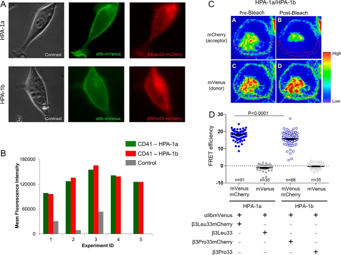
Figure 4.
Transient expression of the complete αIIbβ3 receptor in HEK293 cells. A, phase-contrast and fluorescence microscopy images of a representative HEK293 cell transfected with αIIbmVenus and β3Leu33mCherry plasmids (upper panel) and a representative HEK293 cell transfected with αIIbmVenus and β3Pro33mCherry plasmids (lower panel). B, flow cytometric analyses of αIIbβ3 (CD41), expressing either isoform Leu33 (HPA-1a) or Pro33 (HPA-1b), performed 48 h after transfection in five independent experiments. Of note, the transfectants displayed less than 10% difference in αIIbβ3 expression of either Leu33 (HPA-1a) or Pro33 (HPA-1b) isoform. Values represent mean fluorescence intensity after staining of the transfectants with APC-conjugated CD41 antibody, a complex-specific anti-αIIbβ3 antibody. C, FRET-APB measurements in a representative HEK293 cell transfected with αIIbmVenus and β3Leu33mCherry plasmids. D, results of FRET efficiency of fused individual Leu33 (HPA-1a) or Pro33 (HPA-1b) cells and respective donor controls. To determine the efficiency of energy transfer, the fluorescence of mVenus was measured in a defined region of the membrane (red circled) before and after photobleaching of mCherry at 561 nm (39, 40). Details are given under “Experimental procedures.” The error bars indicate mean ± S.E.

