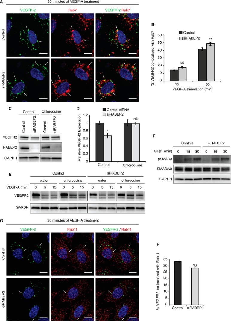
Figure 4.
Assessment of VEGFR2 trafficking mediated by Rab7 and Rab11. A, HUVEC co-stained for internalized VEGFR2 (green) and Rab7 (red) after 30 min of stimulation with 50 ng/ml of VEGF-A165. White arrows point to Rab7+ endosomes containing internalized VEGFR2. DAPI marks cell nuclei in blue; scale bar, 10 μm. B, quantification of co-localization of internalized VEGFR2 and Rab7 after 15 and 30 min of 50 ng/ml of VEGF-A165 stimulation. Quantification was performed using a co-localization plugin of ImageJ in at least 10 independent fields (mean ± S.D., NS, not significant, **, p < 0.01). C, Western blot probed for VEGFR2 of lysates isolated from control and RABEP2 knockdown (siRABEP2) HUVEC grown in complete media and treated with either water (control) or 100 μmol/liter of the lysosomal inhibitor chloroquine for 6 h prior to lysate collection. D, quantification of 3 independent experiments of total VEGFR2 levels normalized to GAPDH in siRABEP2 HUVEC treated with water or chloroquine, relative to control siRNA cells (mean ± S.D., *, p < 0.05). E, Western blot probed for VEGFR2 of lysates isolated from control and RABEP2 knockdown (siRABEP2) HUVEC serum starved for 12 h and then treated with either water (control) or 100 μmol/liter of the lysosomal inhibitor chloroquine for 6 h prior to stimulation for 5 or 15 min with 50 ng/ml of VEGF-A165. GAPDH used as a loading control. F, Western blot of lysates isolated from control and siRABEP2 HUVEC serum starved for 12 h and then stimulated with 1 ng/ml of transforming growth factor β1 (TGFβ1) for 0, 15, and 30 min. Western blots were probed for pSMAD3, a mediator of TGFβ downstream signaling, total SMAD2/3, and GAPDH as a loading control. G, HUVEC co-stained for internalized VEGFR2 (green) and Rab11 (red) after 30 min of stimulation with 50 ng/ml of VEGF-A165. White arrows point to Rab11+ endosomes containing internalized VEGFR2. DAPI marks the cell nuclei in blue; scale bar, 10 μm. H, quantification of co-localization of internalized VEGFR2 and Rab11 after 30 min of 50 ng/ml of VEGF-A165 stimulation. Quantification was performed using a co-localization plugin of ImageJ in at least 10 independent fields (mean ± S.D., NS, not significant).

