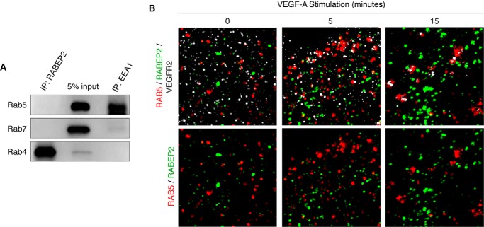
Figure 5.
Assessment of RABEP2 and Rab GTPase interaction. A, Western blot of lysates isolated from HUVEC grown in full media following immunoprecipitation (IP) with antibody against RABEP2 (IP: RABEP2) or EEA (IP: EEA1) and then probed for Rab5, Rab7, and Rab4. 5% input is shown in the middle lane. B, HUVEC infected with adenovirus expressing V5-tagged RABEP2, serum starved overnight, and stimulated for 0, 5, or 15 min with 50 ng/ml of VEGF-A165. Following fixation, cells were co-stained for V5 to visualize RABEP2 (green), RAB5 (red), and VEGFR2 (white) and imaged using SIM with an x-y resolution of 130 nm.

