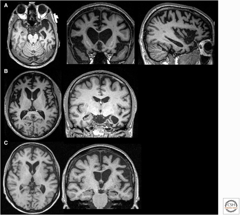
Figure 2.
Magnetic resonance imaging (MRI) in three variants of frontotemporal dementia (FTD). T1-weighted brain MRIs in behavioral variant FTD (bvFTD) (A), semantic variant primary progressive aphasia (svPPA) (B), and nonfluent variant (nfvPPA) (C). (A) A 55-year-old woman with a 4-year history of bvFTD with a score of 27/30 on the mini-mental status examination (MMSE) showing an axial, coronal, and sagittal (right side) MRI with significant bilateral (right more than left) frontal atrophy. (B) A 61-year-old man with svPPA showing symptoms for 1.5 years that included forgetting the names of friends and the names and knowledge of common objects. He also showed difficulty with planning, multitasking, and marked rigidity of daily routines. MRI shows severe left temporal pole atrophy. (C) A 74-year-old man with 2 years of progressive word-finding difficulty, slowed and effortful speech, phonemic paraphasias, and speech apraxia. MRI shows left insular and perisylvian atrophy consistent with nfvPPA. Orientation of coronal and axial MRIs are radiologic. (Images courtesy of Dr. David Perry.)

