Figure 1.

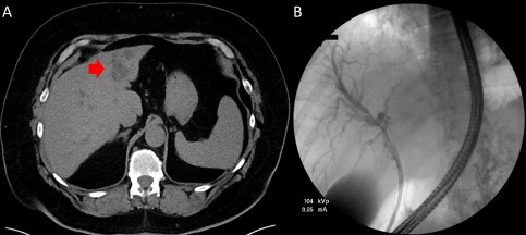
Initial images of the liver. (A) Initial CT abdomen without contrast. Ill‐defined 3.2 × 2.8‐cm mass‐like density in the left hepatic lobe (red arrow). (B) Initial ERCP with stent placed into the left intrahepatic biliary tree.

Initial images of the liver. (A) Initial CT abdomen without contrast. Ill‐defined 3.2 × 2.8‐cm mass‐like density in the left hepatic lobe (red arrow). (B) Initial ERCP with stent placed into the left intrahepatic biliary tree.