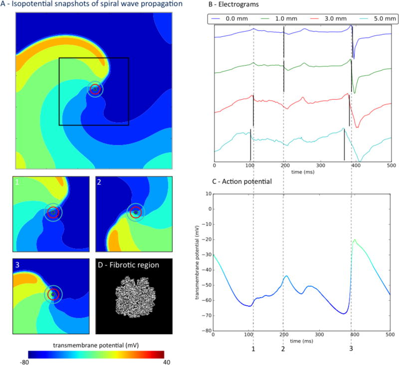
Figure 3.
Atrial fibrillation electrograms poorly represents timing of local activity in the presence of substantial fibrotic remodeling and for larger electrodes. A) Spiral wave propagates through fibrotic tissue, with electrodes of varying diameters marked by concentric circles. Panels 1–3 are isopotential snapshots at marked times; Panel D indicates fibrotic region. B) Atrial fibrillation electrograms generated by different electrodes sizes, with black lines marking minimum dV/dt. Large electrodes (3, 5mm) record a non-local deflection of the wave front that is passing nearby at time 1, whereas small electrodes (0, 1mm) record a deflection that is not a complete activation at time 2 (Panel A, time 2, light blue). C) Action potential at the electrode site is complex due to the interaction of fibrotic and remodeled tissue.

