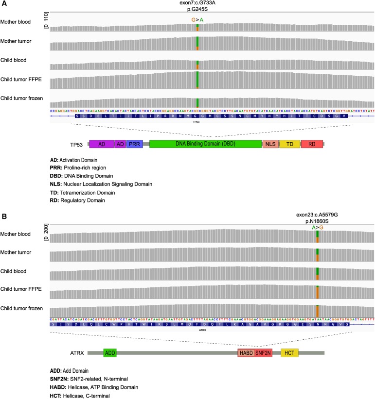
Figure 2.
Both the mother and the child carried a germline mutation in both TP53 and ATRX genes. (A) The sequencing coverage and TP53 allele fraction information in blood and tumor samples as visualized in integrative genomics viewer (IGV). The green color represents the variant allele. Copy-neutral LOH was observed in all the tumor samples. The sequencing coverage range is indicated in square brackets. The exon, transcript base, and amino acid information in the major transcript (NM_000546) are marked above the figure. (Bottom) TP53 mutation is located at the DNA-binding domain. (B) The sequencing coverage and ATRX allele fraction information in blood and tumor samples as visualized in IGV. The orange color represents the variant allele. Copy-neutral LOH was observed in both AT/RT tumor samples. The exon, transcript base, and amino acid information in the major transcript (NM_000489) are marked above the figure. (Bottom) ATRX mutation is located at the SNF2N domain.

