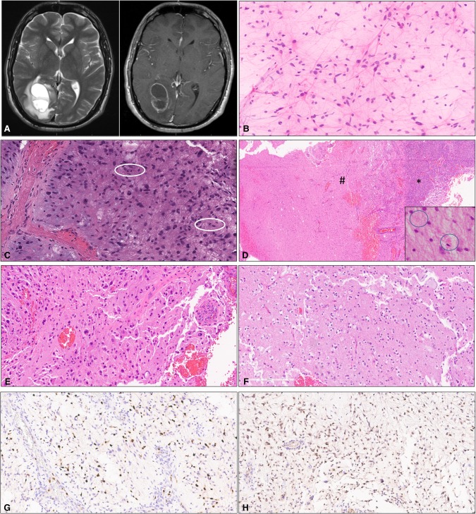
Figure 1.
(A) Axial T2-weighted MRI sequence (left) and T1-weighted MRI sequence with contrast (right) showing the presence of a complex lesion in the right occipital lobe with ring enhancement. (B) Cytologic smear preparation showing neoplastic astrocytes with long, delicate bipolar cytoplasmic processes (pilocytic morphology). (C) Mitotic activity and myxoid background. (D) Arcade of vascular proliferation (*) and relatively sharp demarcation with adjacent brain parenchyma (#). Rare eosinophilic granular bodies (EGBs) were present (inset). (E) Hypercellular brain parenchyma with pleomorphic tumor cells. (F) Tumor areas with myxoid appearance. (G) Ki67 antigen (MIB1) immunostain showing elevated labeling in the region with anaplasia. (H) Expression of p53 protein by tumor cells in the region with anaplasia.

