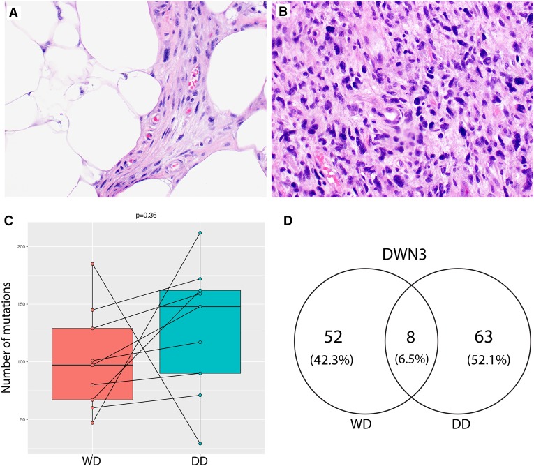
Figure 1.
Whole-exome profiling of concurrent well-differentiated (WD) and dedifferentiated (DD) liposarcomas. (A,B) Representative hematoxylin and eosin stains of liposarcomas used in this study: WD and DD. (C) Boxplots of the total numbers of somatic mutations called by MuTect when comparing tumors with their matched normal samples. Only fresh frozen cases of WD (left) and DD (right) liposarcomas were included here. A paired t-test comparing the somatic mutation burdens between WD and DD liposarcomas was not significant (P = 0.36). (D) Venn diagram example showing the number of somatic mutations in patient 3 (DWN3) as called by MuTect.

