Fig. 1.

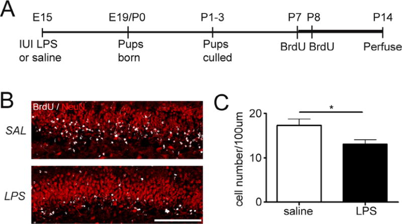
Exposure to intrauterine inflammation reduces early postnatal neurogenesis. (A) Schematic of experimental design. Pregnant dams were given intrauterine injections of lipopolysaccharide (LPS) [50 μg] or saline on embryonic day 15. Pups delivered at term were culled [5 pups/dam] and given injections of bromodeoxyuridine [BrdU, 75 mg/kg; s.c.] at postnatal days 7 and 8 (P7–P8) to label actively proliferating cells. Mice were transcardially perfused with paraformaldehyde at postnatal day 14 (B). Confocal maximum projection images of BrdU (white) and Neuronal Nuclei (NeuN, red) co-labeling in the dentate gyrus of mice exposed to intrauterine saline or LPS and sacrificed at P14. Scale bar = 100 μm. (C) Quantification of BrdU/NeuN density in the granule cell layer at P14. LPS: n = 10; saline: n = 8. *p<0.05. Data represent the mean ± SEM.
