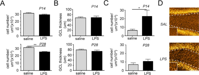
Fig. 2.

Intrauterine inflammation leads to decreased total neuronal density and transient migration abnormalities in the dentate gyrus. Quantification of granule cell density in the granule cell layer (GCL) (A), granule cell layer thickness (B), and ectopic granule cell density in the hilus (C) at postnatal days 14 and 28. *p < 0.05. Data represent the mean ± SEM. (D) Confocal maximum projection images of the granule cell-specific marker Prospero Homeobox 1 (Prox1) in the dentate gyrus of mice exposed to intrauterine saline or lypopolysaccharide (LPS) and sacrificed at P14 (LPS: n = 10; saline: n = 8). Dotted lines depict the granule cell layer (GCL)/hilar border. High magnification images depicted on the bottom left were taken from the center of the hilus. Scale bar = 100 μm, magnified panel = 10 μm.
