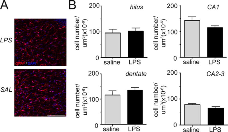
Fig. 6.

Intrauterine inflammation has no effect on hippocampal astrocyte density. (A) Confocal maximum projection images of the astrocyte-specific marker Glial Fibrillary Acidic Protein (GFAP, red) in the Cornu Ammonis 1 (CA1) hippocampal region of mice exposed to intrauterine saline or lipopolysaccharide (LPS) and sacrificed at postnatal day (P14). Cell nuclei are labeled with 4′,6-diamidino-2-phenylindole (DAPI) counterstain (blue). Scale bar = 100 μm. (B) Quantification of astrocyte density in the dentate gyrus, hilus, CA1, and CA2-3 regions at P14. Data represent the mean ± SEM. P14: LPS, n = 10; saline, n = 8.
