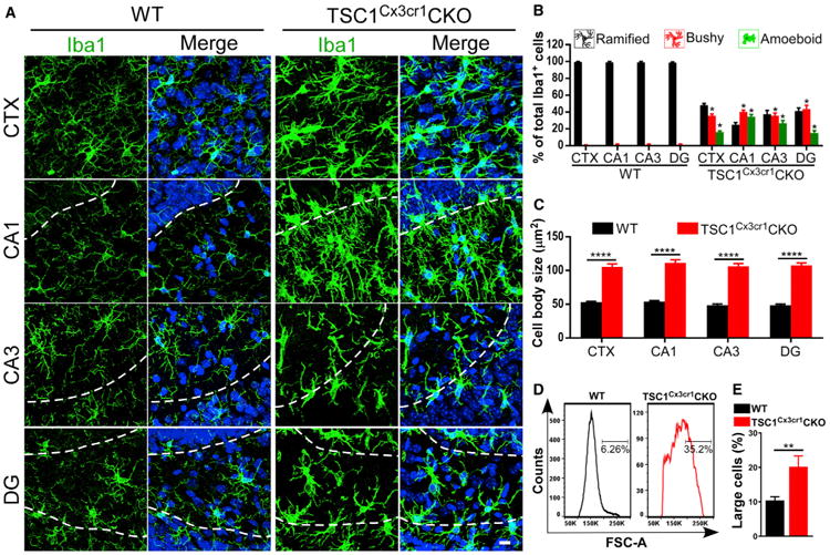
Figure 1. Reactive-like Morphology of Microglia in TSC1Cx3cr1CKO Mice.

(A) Confocal images acquired from cortex (CTX), hippocampal CA1 and CA3, and the dentate gyrus (DG) of Cx3cr1-cre (hereafter referred to as wild-type [WT]) and TSC1Cx3cr1CKO mouse brains. Dashed lines indicate the borders of the CA1 and CA3 pyramidal layers and the dentate granular layers. Scale bar, 10 μm.
(B) Percentage of microglia displaying a ramified, bushy, or amoeboid morphology in the CTX and hippocampus of WT (n = 6; 3 males and 3 females) and TSC1Cx3cr1CKO (n = 7; 3 males and 4 females) mice.
(C) Quantification of microglial cell-body size in WT (n = 6; 3 males and 3 females) and TSC1Cx3cr1CKO (n = 6; 3 males and 3 females) mice.
(D) FACS analysis of cell sizes of microglia (gated as CD11b+/CD45low & Int) in WT and TSC1Cx3cr1CKO mice.
(E) Quantification of the population of microglia with larger cell sizes in WT (n = 4; 2 males and 2 females) and TSC1Cx3cr1CKO (n = 4; 2 males and 2 females) mice. Data are presented as mean ± SEM t test. *p < 0.05, **p < 0.01, and ***p < 0.0001. See also Figure S1.
