
Figure 3. Prolonged VO2 recovery delay is associated with impaired hemodynamic response to exercise.
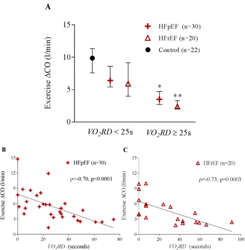
A) Cardiac output augmentation during exercise for the controls, HFpEF, and HFrEF groups is depicted as median with IQR. HFpEF and HFrEF groups are stratified by the median HF VO2RD (25s). (* indicates p=0.0015 between HFpEF < 25s and HFpEF ≥25s and ** indicates p=0.003 between HFrEF < 25s and HFrEF ≥25s. A scatter plot of cardiac output augmentation during exercise versus VO2RD for B) HFpEF and C) HFrEF. Spearman rank correlation is included.