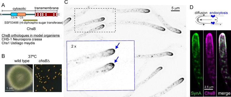
Fig 1. Polarization of ChsB.
(A) domain organization of ChsB. CS-N, Chitin_synth_1N (PF08407); CS, Chitin_synth_1 (PF01644); the C-terminal transmembrane region includes 7 predicted helices (gray boxes). (B) Growth phenotype of chsBΔ microcolonies compared to the wt; plates incubated for 3 days at 37°C. (C) Subcellular localization of endogenously tagged GFP-ChsB. Arrows in the magnified inset indicate the Spitzenkörper (SPK). The image is a MIP of a deconvolved z-stack. (D) The synaptobrevin homologue SynA and ChsB strictly colocalize in the apical crescent, besides the SPK. Images are MIPs of deconvolved z-stacks. The scheme shows an interpretation of endocytic recycling.

