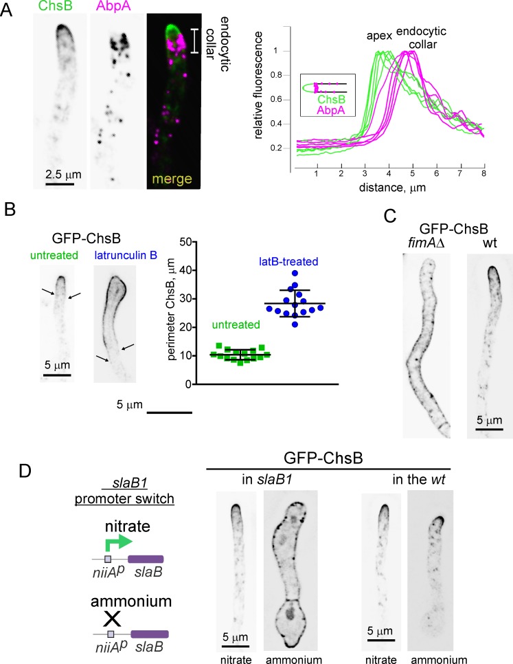
Fig 2. Endocytosis required to maintain ChsB polarity.
(A) The basal limit of the GFP-ChsB apical crescent coincides with the position of the endocytic collar labeled with mCh-AbpA (actin binding protein1). Right graph, linescans along the longitudinal hyphal axis for the green (ChsB) and red (AbpA) channels. (B) Latrunculin B (100 μM) treatment facilitates the diffusion of GFP-ChsB to plasma membrane regions located far away from the apex. Arrows indicate the limits of the PM region occupied by ChsB in untreated and treated examples. The perimeters of the regions occupied by GFP-ChsB in the PM of treated and untreated hyphae are plotted on the right (n = 15 tips). Error bars indicate S.D. The two populations, which passed normality tests, are significantly different (P < 0.0001) in an unpaired t-test with Welch’s correction. (C) Localization of ChsB in a fimAΔ hypha compared to the wt. (D) Scheme: slaB1 drives expression of SlaB under the control of the nitrite reductase promoter (niiAp); Images show the localization of ChsB in a strain carrying the conditional expression allele slaB1 as the only source of SlaBSla2 and its comparison with the wt. slaB1 drives expression of this key endocytic regulator on nitrate as N source but not on ammonium. The germlings derived from conidiospores continuously cultured on medium containing nitrate or ammonium, as indicated. All images represent MIPs of deconvolved z-stacks.

