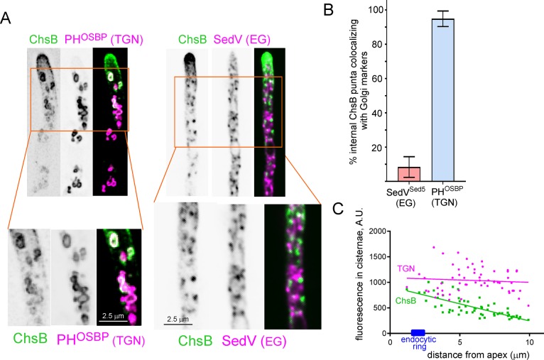
Fig 4. ChsB localizes to the tip-proximal cisternae of the TGN.
(A) colocalization of internal puncta containing GFP-ChsB with the TGN marker mRFP-PHOSBP, and absence of colocalization with the early Golgi marker mCh-SedVSed5 (syntaxin 5). Note the characteristically fenestrated structures of the TGN puncta in the left panels. (B) Quantitation of internal ChsB structures that contain Golgi markers for 178 puncta in n = 10 mRFP-PHOSBP hyphae and 122 puncta in n = 11 mCh-SedVSed5 hyphae. Error bars indicate mean ± SD. The two datasets were significantly different (P< 0.0001) in an unpaired t-test. (C) Plot of fluorescence intensities in the PHOSBP and the ChsB channels vs. distance to the apex. Data of n = 60 TGN cisternae were pooled from 6 hyphae. The fluorescence of ChsB negatively correlates with the distance to the apex (Pearson’s r = -0.748, P = 6E-12) whereas that of PHOSBP does not (r = -0.08, P = 0.53).

