Fig 5.
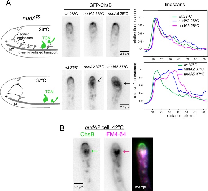
Recycling of ChsB from endosomes necessitates dynein (A) Left, schematics summarizing the rationale of these experiments. Middle images: localization of GFP-ChsB in hyphal tips of the wt and of strains carrying ts mutations in nudA encoding the dynein heavy chain, before and after shifting cells from 28°C to 37°C. Note the aggregate of ChsB in the nudA mutants at 37°C (see scheme), which forms apparently at the expense of the signal in the apical dome and the SPK (which is not detectable in the mutants at 37°C). Right, linescans of the ChsB channel for the hyphae displayed in the images (1 px, 0.103 μm). (B) Hyphal tip of a nudA2 cell shifted to 42°C, stained with the endocytosed membrane tracker FM4-64.

