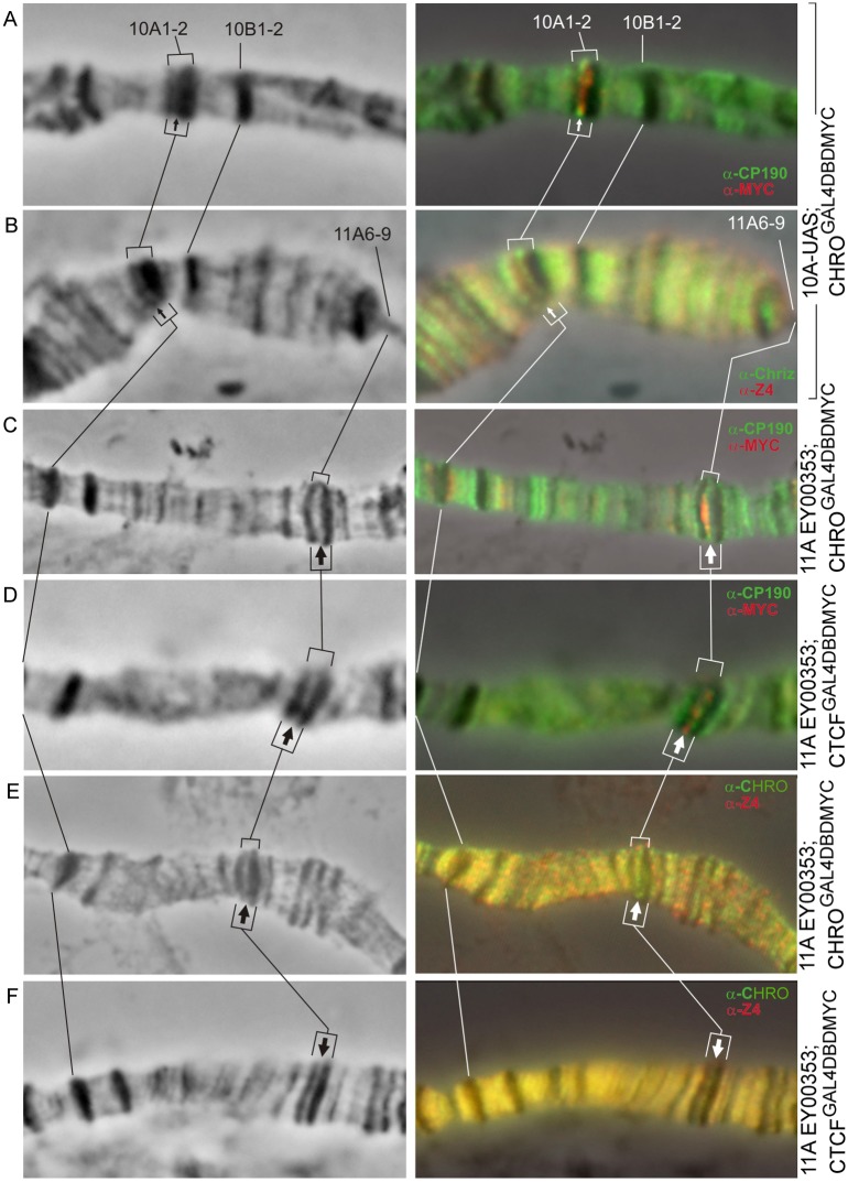
Fig 7. Immunodetection of insulator proteins in the decompacted regions formed within the bands 10A1-2 (A, B) and 11A6-9 (C–F).
Left column: 10A1-2 and 11A6-9 region of the X chromosome (phase contrast), right column: overlay of phase contrast and immunostaining data. Arrows indicate the position of decompaction zone within 10A1-2 (thin arrows) and 11A6-9 (thick arrows) bands.

