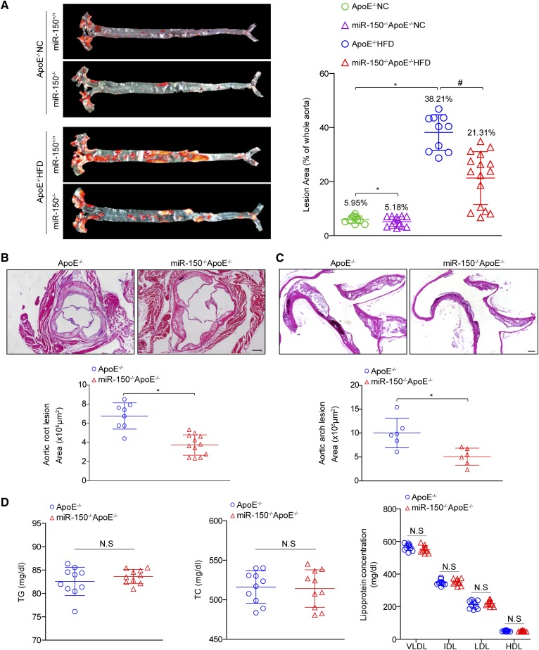
Fig. 2.
miR-150 ablation protects mice from atherosclerosis. A: Oil Red O staining of atherosclerotic lesions in the entire aorta in ApoE−/− and miR-150−/−ApoE−/− mice treated with NC or a HFD for 28 weeks. Quantitative lesion data are shown in the right panel (n = 10–16). *P < 0.05 versus ApoE−/− NC group; #P < 0.05 versus ApoE−/− HFD group. B, C: H&E staining for the morphology of the plaques in the aortic roots (B) and ascending aortic arches (C) of ApoE−/− and miR-150−/−ApoE−/− mice treated with a HFD for 28 weeks (n = 6–12). Scale bar = 200 μm. *P < 0.05 versus ApoE−/− group. D: The lipid metabolism parameters of ApoE−/− and miR-150−/−ApoE−/− mice (n = 10 each group). Not significant (N.S) versus ApoE−/− littermates. TC, total cholesterol; TG, triglyceride.

