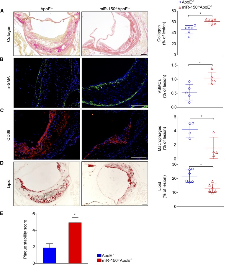
Fig. 3.
miR-150 deficiency increases plaque stability. A–D: Cross-sections of aortic roots from ApoE−/− and miR-150−/−ApoE−/− mice were stained with picrosirius red to assess collagen deposition (A) and α-smooth muscle actin (α-SMA) expression (green) to determine SMC compositions (B). The tissues were also stained with CD68 (red) to assess macrophage infiltration (C) and Oil Red O to assess lipid accumulation (D). The quantitative results for each image are shown in the right panels. E: The assessment of plaque stability score in ApoE−/− and miR-150−/−ApoE−/− mice (n = 4–6). Scale bar = 100 μm.*P < 0.05 versus ApoE−/− group.

