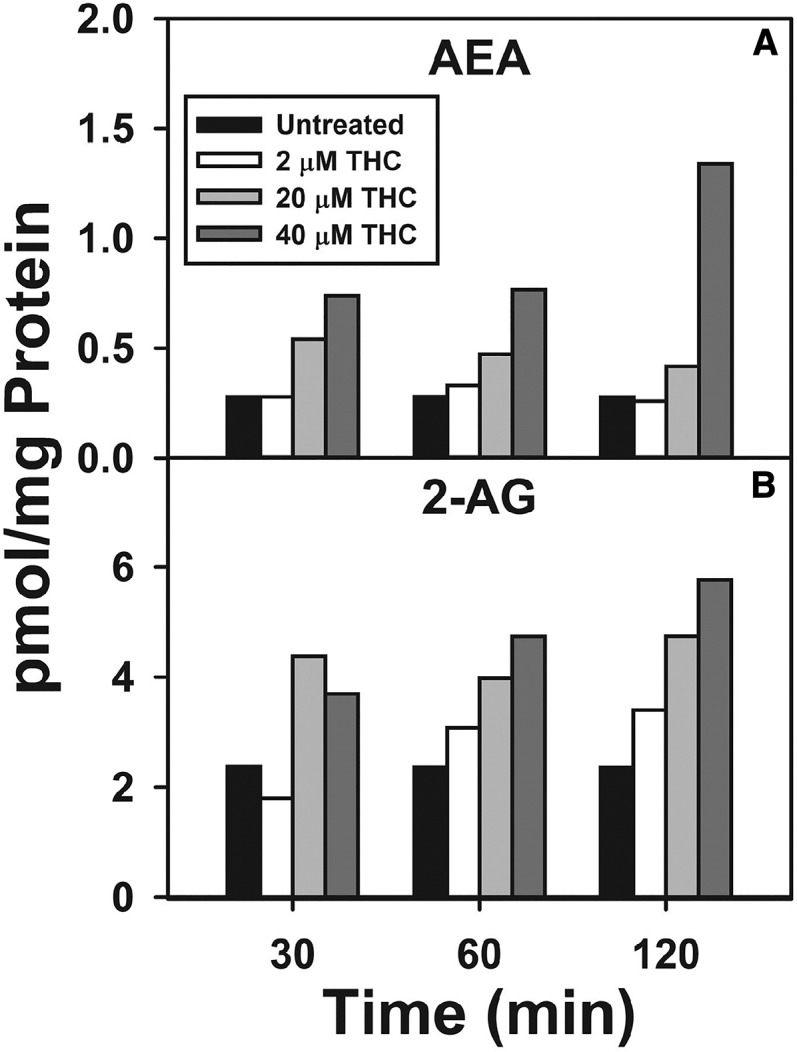
Fig. 1.

Time and concentration dependence of Δ9-THC impact on hepatocyte levels of AEA and 2-AG. Cultured primary hepatocytes were isolated from WT mice, plated on culture dishes, and incubated with increasing Δ9-THC (2–40 μM) for increasing time (0–2 h), as described in the Materials and Methods. Lipids were then extracted followed by analysis and quantitation by LC-MS as described in the Materials and Methods. AEA (A) and 2-AG (B) (n = 1).
