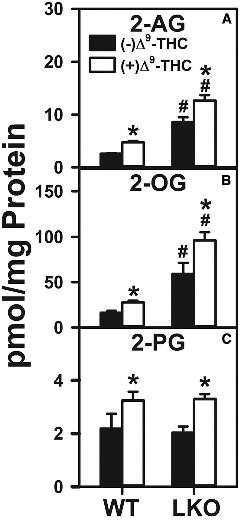
Fig. 4.

Effect of Δ9-THC and Fabp1 gene ablation on hepatocyte levels of 2-AG and non-ARA-containing 2-MGs. Hepatocytes were isolated from WT or LKO mice, plated on culture dishes, and incubated with Δ9-THC (20 μM) for 1 h, as described in the Materials and Methods. Lipids were then extracted followed by 2-AG and 2-MG analysis and quantitation by LC-MS, also as described in Materials and Methods. 2-AG (A), 2-OG (B), and 2-PG (C). Values represent the mean ± SEM, n = 4–6. #P < 0.05 versus WT in the same treatment groups; *P < 0.05 versus untreated of the same genotype.
