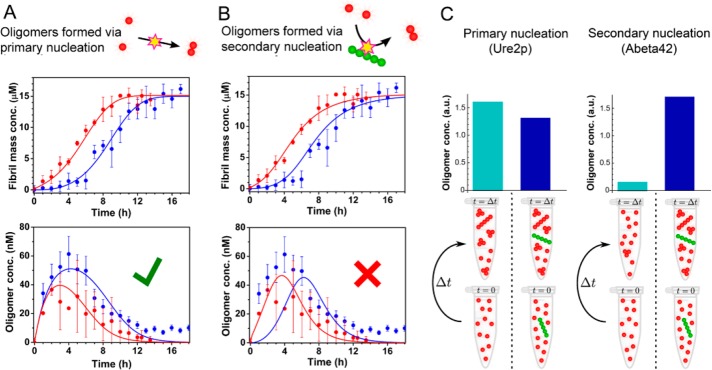
Figure 2.
Absence of a fibril-catalyzed secondary nucleation process for Ure2. (A, B) Ensemble aggregation kinetics of 15 μM unlabeled Ure2-S68C monitored by ThT fluorescence under unseeded (blue) or seeded (red) conditions (upper panels). Ure2 oligomers were then detected under unseeded (blue) or seeded (red) conditions by confocal single molecule FRET (lower panels). The incubation conditions were the same as in Figure 1. (A) The data fit well to a model that generates oligomers during primary nucleation. (B) A model that generates oligomers during secondary nucleation cannot fit the data. (C) The presence of seeds (right-hand columns) drastically increases the concentration of Aβ42 oligomers measured at a single time point in the lag phase of an Aβ42 aggregation experiment52 (right panel), but do not increase the production of Ure2 oligomers in this study (left panel), indicating fundamentally different mechanisms of oligomer formation for these two systems.

